Radiographs For Neck Pain May Include X-Rays, Mri Or Ct Scan
For mechanical neck pain without neurological findings, most doctors find it reasonable to wait about 6-8 weeks before taking x-rays or other radiographs for neck pain. Imaging studies are not always necessary and with mechanical pain without any red flags or warning signs, it is reasonable to proceed with conservative neck pain treatment.
If you see a Chiropractor, x-rays are usually taken as part of the initial evaluation. If the neck pain persists beyond a 6-8 week period, most medical doctors will find it helpful to obtain x-rays and determine if further studies are needed.
As mentioned previous, trauma, severe symptoms with unrelenting pain which is getting worse, fever, chills, sweats or recent and significant weight loss may indicate a pathological condition for which radiologic examinations should not be delayed.
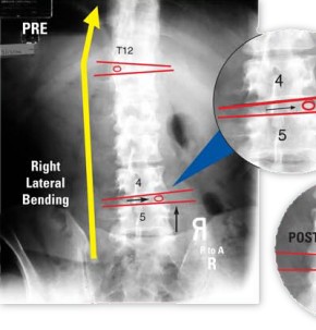
Conventional radiographs would include x-rays that take pictures of the front/back and side of the neck called anteroposterior and lateral views. Flexion and extension (forward and back bending) views may also be taken to look for instability or aberrant motion of the spinal segments involved in these neck motions.
 The x-rays are also evaluated for curvature of the neck. The normal curve of the neck formed by the spinal bones is a backward c-shaped curve called the lordosis like the picture. Loss of this curve may be structural, due to advancing disc degeneration and loss of the disc height, or it may be due to muscle spasm from pain.
It should be noted that discs cannot be imaged on plain x-rays. The extent of degeneration is seen in the associated changes of the bones, which may include loss of disc height, bone spurs, bone irregularities and increased signaling or whiteness at the ends of the bones above and below the disc space.
MRI or magnetic resonance imaging is useful in viewing disc problems to determine if they are dehydrated or diseased, where they appear black. Severely degenerated discs may show accumulating gas in disrupted areas of the degenerated discs and this is called a “vacuum disc sign”.
MRI is also good for determining if there are any tears in the outer part of the disc called the annulus which may cause pain or be a signs that the degenerative process is underway.
CT scans (Computed Tomography) can give more information on the amount of bone destruction in cases of trauma, infection or tumors, however, they are not very useful or practical for mechanical neck pain evaluation. In cases with radiculopathy involving the nerve root, CT scans may be helpful in viewing the opening for the nerves or the bony tunnel from which nerves exit between 2 bones called “foramina”.
After a review of radiographs for neck pain, the next part focuses on cervical strain including whiplash, which is the most common cause of mechanical or axial neck pain.
Mechanical Neck Pain | Features Neck Pain | Cervical Strain | Cervical Degenerative Disc Disease | C4 Radiculopathy | Failed Neck Surgery | Rheumatoid Neck Disorders | Shoulder Problems | Mechanical Neck Pain Summary

