
What Is An MRI Scan?
 An MRI scan is a test to look into the neck & back, which doesn’t use any radiation. Instead of radiation, they use magnetic & radio waves, creating computer generated pictures. The pictures or images produced by an MRI can show different layers of the spine and are able to show problems with soft tissues, like muscles, ligaments, spinal discs and nerves. If you are scheduled for scanning, you should know that it is the most common test for looking into the spine other than a standard x-ray.
What Does An MRI Scan Show?
An scan can show clear details of the spine. It provides pictures that show the condition of discs, ligaments, muscles and bones in much greater detail than an x-ray. It can show early signs of disc degeneration by focusing on the central part of the disc called the nucleus. Observing loss of water in the nucleus is an early sign of disc degeneration. It can show details of degeneration that diagnose specific forms most related to back pain like Modic changes.
It can also show the conditions of the joints, which are frequently involved in facet joint arthritis. By looking into the spine, a scan can show areas where the spinal canal has become narrowed, a condition called spinal stenosis. It can also show if there is a herniated disc in the neck or back, and can tell how big it is and if it is pressing on the nerve or parts of the spinal cord.
What Does It Not Show?
It shows most structures very well, however, an x-ray can show bone in finer detail and, many doctors that need to look at destruction of bone from tumors or infection and fractures will choose a CT Scan.
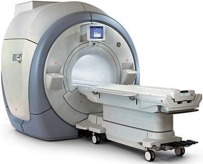
How Is An MRI Scan Done?
During the scan, you will lie down on a table that will slide into the main machine that resembles a round tunnel. The technician will make sure you are comfortable by using pillows and, often you can listen to music. While the scan is being done, many pictures are taken. During the process, the technician carefully monitors everything and can communicate with you. Often, you will be given a hand held button to press if you experience any problems. The scanner does not provide a lot of room, so some people may get a bit claustrophobic. If this is the case, a mild sedative may be given to ease the anxiety. Open MRI scan machines minimize this with a more open feel.
How Long Does An MRI Scan Take?
It usually takes anywhere from 30 to 60 minutes.
Are They Noisy?
Yes, but this depends on the machine. Some are more noisy than others. It can be a humming and banging type of sound. Headphones with music are usually provided for the noisier machines, while background music is enough for the quieter ones. The noise is just when the scanning is being done. In between scans, it is quiet. The technician may say, “OK, This scan will take 10 minutes”, as the settings are prepared.
Contraindications
Although there is no problems from the magnetic waves regarding exposure, you should not have one if there are any metal objects in your body. Mostly from past surgeries, these metal objects can move or come loose in the magnetic field during the scan. If you are not sure, this needs to be addressed. Sometimes, x-rays can be taken if there is a suspicion of metal in the body. The facility will have a list of objects that you need to verify you do not have, even permanent eyeliner. You should not wear any jewelry or clothes with metal clips, zippers or buttons. Wearing a natural fiber sweatsuit is a good idea.
How Much Does It Cost?
The costs vary and can be anywhere from 1 to 3 thousand dollars, in general. The variables are many, since insurance coverage also varies as well as special studies done with contrast or other special parameters. The technician fee is associated with the test itself, and there is also a professional fee for a radiologist to read the scans and write a report. There may be 2 separate costs or it may be included in one price depending on the facility.
Is It Necessary?
A 2021 study in the International Journal for Quality in Health Care is an attempt to reduce usage of MRIs. For mechanical back pain, the recommendations for MRI are: (with symptoms persisting or worsening despite conservative management for at least 6 weeks) Low back pain for at least 6 months (Disc Pain & Facet Joint Pain). Radiculopathy for at least 6 weeks (Compressed Nerve Pain). Spinal stenosis symptoms for at least 6 weeks (Neurogenic Claudication). Noted: Patient has above mentioned indications, but with contraindication’s like claustrophobia, heart pacemaker, intracranial metal clips… – CT instead.
They state, “Please note that imaging tests like X- rays, CT scans and MRIs are not helpful for recovery or management of acute or recurring low back pain unless there are signs of serious pathology.”
While studies may be abused and this should be discouraged, attempts to reduce cost – or payment may not be advisable in many conditions and may foster chronic pain, psychological concern, and delay targeted treatment to a pathological generator other than “serious pathology”.